
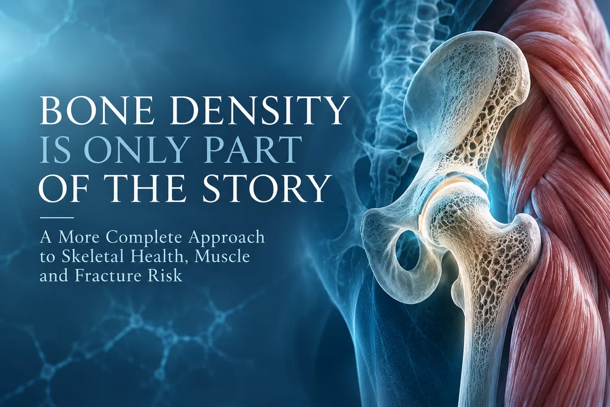
Bone Density Is Only Part of the Story
A More Complete Approach to Skeletal Health, Muscle and Fracture Risk
Osteoporosis is frequently discussed as a disorder of low bone mineral density, but bone mineral density represents only one component of skeletal strength. The clinically meaningful outcome in osteoporosis is fragility fracture, and fracture susceptibility reflects the interaction of bone mass, bone architecture, tissue properties, remodeling activity, muscle function, mechanical loading, fall risk and numerous clinical variables.
Dual energy X ray absorptiometry, commonly referred to as DXA or DEXA, remains the clinical standard for measuring bone mineral density and is fundamental to the diagnosis and management of osteoporosis. The International Society for Clinical Densitometry distinguishes, however, between the use of bone mineral density for diagnostic classification and its use as one component of fracture risk assessment. This distinction is important because a T score is an exceptionally useful measurement, but it is not a direct measurement of the mechanical strength of bone. (ISCD-)
At the Institute for Human Optimization, our approach to skeletal health begins with this distinction. We do not view additional measurements as replacements for established diagnostic tools. Instead, we evaluate different components of the musculoskeletal system according to the clinical question being asked. Bone density, skeletal muscle mass, body composition, metabolic health, physical function and bone remodeling describe different aspects of the same biological system.
What DEXA Measures
DXA measures areal bone mineral density, expressed in grams per square centimeter. In postmenopausal women and men aged 50 years and older, T scores derived from appropriate skeletal sites are used for densitometric classification of osteoporosis. A T score of −2.5 or below at the femoral neck, total hip, lumbar spine or, under appropriate circumstances, the 33 percent radius can establish the diagnosis.
This threshold has considerable clinical value, but it should not be interpreted as a biological boundary between strong and weak bone. Skeletal fragility exists along a continuum. The International Society for Clinical Densitometry specifically notes that diagnostic classification and fracture risk assessment are distinct concepts and that individuals with low bone mass are not necessarily at high fracture risk. (ISCD -)
The reason is partly mechanical. DXA measures mineral density projected across a two dimensional area. It does not directly measure cortical porosity, trabecular connectivity, three dimensional geometry, material properties or the amount of force necessary to cause structural failure.
Bone strength is therefore influenced by bone quantity, but it is not defined by bone quantity alone.
Bone Quality and Architecture
Bone is a highly organized composite tissue. Its ability to withstand mechanical stress depends on the amount of mineralized tissue present as well as the distribution and organization of that tissue.
Cortical thickness and porosity influence structural integrity. Within trabecular bone, the number, thickness, orientation and connectivity of trabeculae contribute to mechanical competence. Bone geometry determines how forces are distributed through the structure, while tissue composition, mineralization and accumulated microdamage influence its material properties.
This helps explain why two individuals with similar bone mineral density measurements may not have identical fracture susceptibility.
Additional techniques can interrogate some of these characteristics. Trabecular Bone Score, for example, is derived from lumbar spine DXA images and provides information related to trabecular microarchitecture that is not fully captured by bone mineral density alone. Current ISCD positions recognize associations between TBS and vertebral, hip and major osteoporotic fracture risk in appropriate populations and support its use in adjusting fracture risk estimates. Importantly, ISCD also states that TBS should not be used independently to determine treatment. (ISCD -)
This is representative of how we think about advanced phenotyping more broadly: complementary measurements can refine clinical characterization without displacing validated foundational measurements.
The Muscle Bone Unit
A comprehensive discussion of skeletal aging also requires consideration of skeletal muscle.
Bone and muscle are functionally interconnected tissues. Muscle contraction generates mechanical forces that are transmitted to bone, and the skeleton adapts to mechanical loading through mechanosensitive cellular pathways. Osteocytes detect changes in mechanical strain and participate in signaling pathways that regulate the activity of osteoblasts and osteoclasts. The skeletal response to exercise therefore involves considerably more than simply supporting additional body weight.
This relationship becomes particularly important with aging. Progressive reductions in physical activity, resistance exercise, muscle mass and muscle strength can occur at the same time that bone mass and structural integrity are declining. The coexistence of osteoporosis and sarcopenia is increasingly described as osteosarcopenia, reflecting the biological and clinical overlap between deterioration of the skeletal and muscular systems.
The implications extend beyond bone remodeling. Reduced muscle strength can impair balance, mobility and the ability to recover from perturbations, increasing the probability of a fall. Once a fall occurs, the magnitude and direction of the resulting load interact with skeletal strength to determine whether a fracture occurs.
For this reason, resistance exercise is not merely a fitness recommendation added to osteoporosis care. Progressive resistance and appropriately prescribed weight bearing exercise provide mechanical stimuli relevant to both muscle and bone while also addressing strength and physical function.
The clinical objective is therefore broader than preserving bone mineral density. Maintaining the musculoskeletal system requires preserving the tissue that loads the skeleton and the physical capacity that protects it.
Why We Measure Body Composition at the Institute
This muscle bone relationship is one reason body composition is incorporated into our evaluation at the Institute for Human Optimization.
We use InBody analysis in office to characterize body composition, with particular attention to skeletal muscle mass, segmental lean mass and visceral adiposity. These measurements are not intended to diagnose osteoporosis, nor do we use InBody as a substitute for DXA. Their purpose is to characterize additional components of the patient’s metabolic and musculoskeletal phenotype.
Skeletal muscle mass provides useful information about the amount and distribution of lean tissue, particularly when followed longitudinally. Interpretation should extend beyond muscle quantity alone, however. Muscle mass, muscle strength and physical performance are related but distinct characteristics. A complete assessment may therefore incorporate body composition alongside exercise history, strength, functional capacity and other clinical measures when appropriate.
This becomes particularly relevant when a patient has been losing weight. Reduction in total body mass does not reveal how much of that change came from adipose tissue versus lean tissue. In the context of aging and skeletal health, preservation of skeletal muscle can be clinically important.
We also follow visceral adiposity because it provides information about metabolic phenotype that cannot be inferred reliably from body weight or body mass index alone. Visceral adipose tissue is biologically active and has established relationships with cardiometabolic disease. Its relationship with skeletal health, however, is more complex. Observational studies have reported associations between visceral adiposity and bone outcomes, but these associations differ according to sex, total adiposity and other variables, and Mendelian randomization analyses have not established a straightforward causal relationship between visceral adiposity and bone mineral density or fracture. (PubMed)
We therefore do not interpret visceral fat as an osteoporosis biomarker. We measure it because skeletal health exists within a larger metabolic and body composition phenotype, and visceral adiposity is one clinically relevant component of that phenotype.
Formal Bone Density Assessment
When diagnostic bone density imaging is indicated, we work with established radiology centers in the region, including Charter Radiology, to provide formal DEXA imaging.
Charter Radiology offers DEXA scanning at its Maryland imaging centers, including its Clarksville location. (Charter Radiology)
This allows formal radiologic assessment to remain within the appropriate imaging environment while the results can be interpreted within a broader clinical evaluation. Depending on the individual, that evaluation may include medical and fracture history, medications, endocrine and metabolic factors, nutritional status, body composition, skeletal muscle, exercise exposure and biochemical markers of bone metabolism.
The intention is not to make every evaluation more complicated. It is to avoid asking one measurement to answer a question it was not designed to answer.
Bone Remodeling Adds Another Dimension
Bone is not a static structural material. It is continuously remodeled through coordinated osteoclastic bone resorption and osteoblastic bone formation.
This process can be evaluated biochemically through bone turnover markers. The International Osteoporosis Foundation and International Federation of Clinical Chemistry and Laboratory Medicine have identified procollagen type I N terminal propeptide, or PINP, as a reference marker of bone formation and β isomerized C terminal telopeptide of type I collagen, or β CTX I, as a reference marker of bone resorption.
A 2025 international consensus update reaffirmed these markers and reviewed their evidence for fracture risk assessment and monitoring osteoporosis therapy. Higher bone turnover marker concentrations have been associated with greater fracture risk in postmenopausal women, although their clinical interpretation requires appropriate standardization and context. (PubMed)
Bone turnover markers should not be viewed as biochemical substitutes for DXA. They provide different information. DXA characterizes bone mineral density, whereas turnover markers provide information about the rate of ongoing skeletal remodeling.
This distinction can be clinically useful when the question involves not only the current state of the skeleton but also the biological processes contributing to change.
Longitudinal Assessment
A further advantage of integrating these measurements is the ability to evaluate trajectory.
Serial DXA measurements can determine whether bone mineral density is changing beyond the expected precision error of the measurement. The ISCD supports follow up BMD testing when the result is likely to influence clinical management and emphasizes appropriate facility specific precision assessment when interpreting serial changes. (ISCD -)
Body composition can similarly be followed over time to evaluate changes in skeletal muscle and adiposity. Exercise exposure, physical performance and biochemical markers may provide additional context depending on the clinical situation.
This longitudinal perspective is particularly relevant because skeletal deterioration can develop gradually before the first clinical fracture. A patient may simultaneously experience declining bone density, decreasing muscle mass, reduced resistance exercise and worsening metabolic health. Examining those variables independently can miss the relationship among them.
The appropriate objective is not to generate the largest possible dataset. It is to identify clinically meaningful changes early enough to determine whether intervention is warranted.
A Systems Approach to Skeletal Aging
At the Institute for Human Optimization, we approach bone health as part of a larger musculoskeletal and metabolic system.
DEXA remains foundational when bone mineral density assessment is indicated. Body composition provides information about skeletal muscle and adiposity. Physical performance and resistance training history provide context regarding mechanical loading and functional capacity. Bone turnover markers can characterize aspects of remodeling biology in selected patients. Clinical history, medication exposure, endocrine function, nutritional status and previous fracture history remain essential to interpretation.
None of these measurements should be considered in isolation, and not every patient requires every assessment.
The scientific rationale for integrating them is straightforward: fracture susceptibility is a multifactorial phenotype. Bone mineral density is an important determinant of that phenotype, but skeletal architecture, remodeling, muscle, mechanical loading, metabolic health and fall susceptibility contribute additional information.
A precision medicine approach to skeletal health therefore does not require replacing DEXA or redefining osteoporosis. It requires using validated measurements for the questions they are capable of answering and integrating additional information when it has the potential to meaningfully alter risk assessment, prevention or management.
References
International Society for Clinical Densitometry. 2023 ISCD Official Adult Positions. (ISCD -)
Bhattoa HP, et al. Update on the role of bone turnover markers in the diagnosis and management of osteoporosis: a consensus paper from ESCEO, IOF and IFCC. Osteoporosis International. 2025;36(4):579–608. doi:10.1007/s00198-025-07422-3. (PubMed)
Hu J, et al. Associations of visceral adipose tissue with bone mineral density and fracture: observational and Mendelian randomization studies. Journal of Nutritional Health & Aging. 2022. PMID: 35821143. (PubMed)
International Society for Clinical Densitometry. Clinical Use of Trabecular Bone Score: 2023 Official Positions. (ISCD -)
This article is intended for educational purposes and does not constitute individualized medical advice. Diagnostic testing, exercise prescription and treatment decisions should be based on an individual’s clinical history, examination, risk factors and appropriate clinical guidelines.